

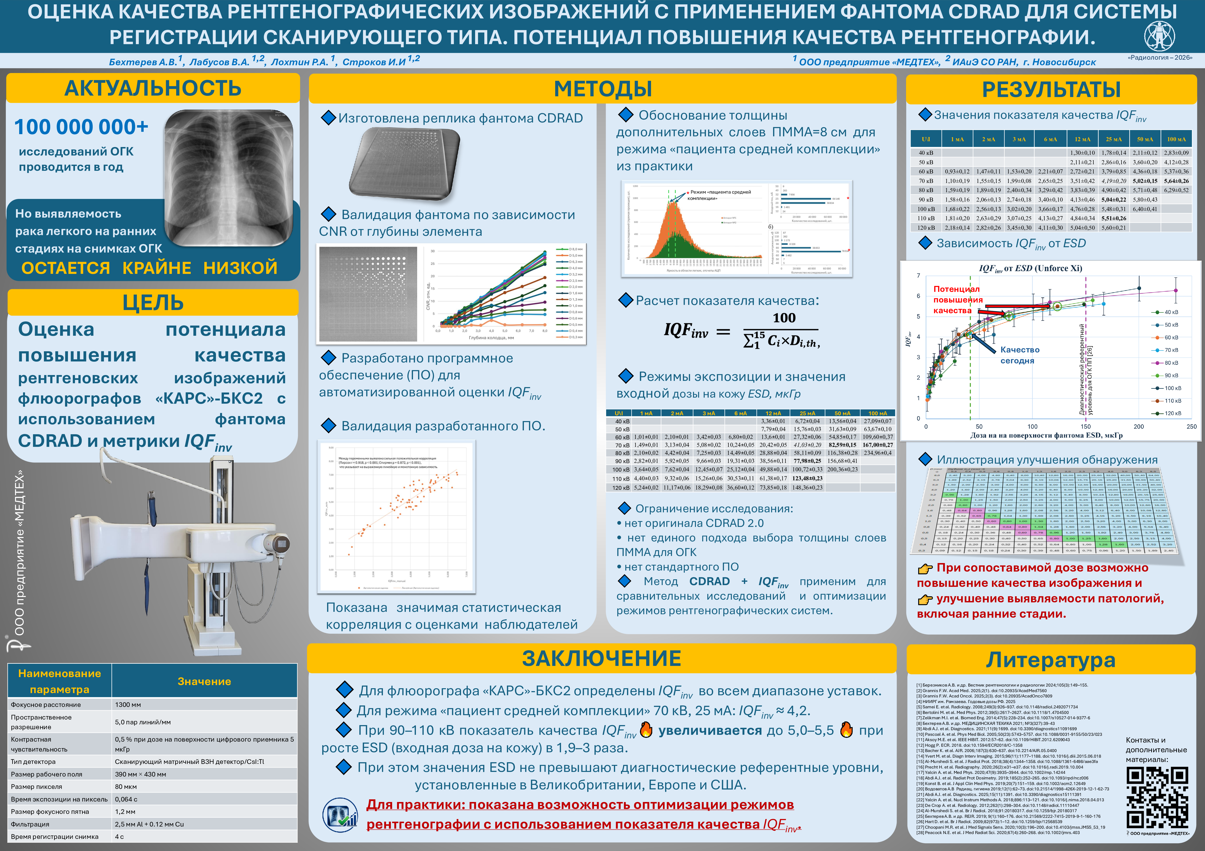
Оценка качества рентгенографических изображений с применением фантома CDRAD для системы регистрации сканирующего типа. Потенциал повышения качества рентгенографии.
Доклад в рамках XX Юбилейного Всероссийского национального конгресса лучевых диагностов и терапевтов «Радиология – 2026» https://radiology-congress.ru/
Список литературы:
- [1] Березников А.В., Шкитин С.О., Тюрин И.Е. Возможности рентгенографии грудной клетки в первичной диагностике рака легкого. Вестник рентгенологии и радиологии. 2024; 105(3): 149–155. https://doi.org/10.20862/0042-4676-2024-105-3-149-155.
- [2] F. W. Grannis, «Why has there been such a long delay in the implementation of population lung cancer screening in the USA?», Academia Medicine, т. 2, вып. 1, мар. 2025, doi: 10.20935/AcadMed7560.
- [3] F. W. Grannis, «Cassandra’s tears: population lung cancer screening in Western Europe», Academia Oncology, т. 2, вып. 3, авг. 2025, doi: 10.20935/AcadOnco7809.
- [4] Справочник: «Годовые эффективные дозы облучения населения Российской Федерации в 2024 году».- СПб.: ФБУН НИИРГ им. П.В. Рамзаева, 2025. – 69 с..
- [5] E. Samei, N. T. Ranger, A. MacKenzie, I. D. Honey, J. T. Dobbins, и C. E. Ravin, «Detector or system? Extending the concept of detective quantum efficiency to characterize the performance of digital radiographic imaging systems», Radiology, т. 249, вып. 3, сс. 926–937, дек. 2008, doi: 10.1148/radiol.2492071734.
- [6] M. Bertolini и др., «A comparison of digital radiography systems in terms of effective detective quantum efficiency», Medical Physics, т. 39, вып. 5, сс. 2617–2627, май 2012, doi: 10.1118/1.4704500.
- [7] М.И Зеликман, С.П. Кабанов, С.А. Кручинин, «Оценка влияния рассеянного в теле пациента излучения на характеристики тракта формирования цифрового рентгеновского изображения», МЕДИЦИНСКАЯ ТЕХНИКА 2013 №5, (281), с. 4-9, doi: 10.1007/s10527-014-9377-6.
- [8] Бехтерев А.В., Котков Р.В., Лабусов В.А., Лохтин Р.А., Пьянов Д.А., Строков И.И., Храмов М.С. DQE и eDQE цифровой рентгенографической системы сканирующего типа «КАРС»-БКС2 // Медицинская техника. №3, 2021. с. 39-43. https://doi.org/10.1007/s10527-021-10103-0.
- [9] A. J. Abdi, B. R. Mussmann, A. Mackenzie, O. Gerke, B. Klaerke, и P. E. Andersen, «Quantitative Image Quality Metrics of the Low-Dose 2D/3D Slot Scanner Compared to Two Conventional Digital Radiography X-ray Imaging Systems», Diagnostics, т. 11, вып. 9, с. 1699, сен. 2021, doi: 10.3390/diagnostics11091699.
- [10] A. Pascoal, C. P. Lawinski, I. Honey, и P. Blake, «Evaluation of a software package for automated quality assessment of contrast detail images—comparison with subjective visual assessment», Phys. Med. Biol., т. 50, вып. 23, сс. 5743–5757, дек. 2005, doi: 10.1088/0031-9155/50/23/023.
- [11] M. E. Aksoy, M. E. Kamasak, E. Akkur, A. Ucgul, M. Basak, и H. Alaca, «Evaluation and comparison of image quality for indirect flat panel systems with CsI and GOS scintillators», в 2012 7th International Symposium on Health Informatics and Bioinformatics, Nevsehir, Turkey: IEEE, апр. 2012, сс. 57–62. doi: 10.1109/HIBIT.2012.6209043.
- [12] P. Hogg, «A comparison of the performance of a 2.4 MP colour monitor and a 5.0 MP monochrome monitor in visualising low contrast detail using the CDRAD phantom», с. 996 words, 2018, doi: 10.1594/ECR2018/C-1358.
- [13] K. Bacher и др., «Image Quality and Radiation Dose on Digital Chest Imaging: Comparison of Amorphous Silicon and Amorphous Selenium Flat-Panel Systems», American Journal of Roentgenology, т. 187, вып. 3, сс. 630–637, сен. 2006, doi: 10.2214/AJR.05.0400.
- [14] M. Yvert, A. Diallo, P. Bessou, J.-L. Rehel, E. Lhomme, и J.-F. Chateil, «Radiography of scoliosis: Comparative dose levels and image quality between a dynamic flat-panel detector and a slot-scanning device (EOS system)», Diagnostic and Interventional Imaging, т. 96, вып. 11, сс. 1177–1188, ноя. 2015, doi: 10.1016/j.diii.2015.06.018.
- [15] S. Al-Murshedi, P. Hogg, L. Lanca, и A. England, «A novel method for comparing radiation dose and image quality, between and within different x-ray units in a series of hospitals», J. Radiol. Prot., т. 38, вып. 4, сс. 1344–1358, дек. 2018, doi: 10.1088/1361-6498/aae3fa.
- [16] H. Precht и др., «Comparison of image quality in chest, hip and pelvis examinations between mobile equipment in nursing homes and static indirect radiography equipment in the hospital», Radiography, т. 26, вып. 2, сс. e31–e37, май 2020, doi: 10.1016/j.radi.2019.10.004.
- [17] A. Yalcin, T. Olgar, T. Sancak, G. K. Atac, и S. Akyar, «Correlation between physical measurements and observer evaluations of image quality in digital chest radiography», Medical Physics, т. 47, вып. 9, сс. 3935–3944, сен. 2020, doi: 10.1002/mp.14244.
- [18] A. J. Abdi, B. Mussmann, A. Mackenzie, B. Klaerke, и P. E. Andersen, «COMPARISON OF RADIATION EXPOSURE TO THE PATIENT AND CONTRAST DETAIL RESOLUTIONS ACROSS LOW DOSE 2D/3D SLOT SCANNER AND TWO CONVENTIONAL DIGITAL RADIOGRAPHY X-RAY IMAGING SYSTEMS», Radiation Protection Dosimetry, т. 185, вып. 2, сс. 252–265, дек. 2019, doi: 10.1093/rpd/ncz006.
- [19] B. Konst, H. Weedon‐Fekjær, и M. Båth, «Image quality and radiation dose in planar imaging — Image quality figure of merits from the CDRAD phantom», J Applied Clin Med Phys, т. 20, вып. 7, сс. 151–159, июл. 2019, doi: 10.1002/acm2.12649.
- [20] Водоватов А.В. Использование тест-объекта «контраст-деталь» для оценки возможности снижения доз облучения пациентов в цифровой рентгенографии органов грудной клетки // Радиационная гигиена. – 2019. – Т. 12, No 1. – С. 62-73. DOI: 10.21514/1998-426Х-2019-12-1-62-73.
- [21] A. J. Abdi, H. Precht, C. B. Outzen, и J. Jensen, «Quantitative Evaluation of Image Quality and Radiation Dose Using Novel Intelligent Noise Reduction (INR) Software in Chest Radiography: A Phantom Study», Diagnostics, т. 15, вып. 11, с. 1391, май 2025, doi: 10.3390/diagnostics15111391.
- [22] A. Yalcin и T. Olgar, «Characterizing the digital radiography system in terms of effective detective quantum efficiency and CDRAD measurement», Nuclear Instruments and Methods in Physics Research Section A: Accelerators, Spectrometers, Detectors and Associated Equipment, т. 896, сс. 113–121, июл. 2018, doi: 10.1016/j.nima.2018.04.013
- [23] A. De Crop и др., «Correlation of Contrast-Detail Analysis and Clinical Image Quality Assessment in Chest Radiography with a Human Cadaver Study», Radiology, т. 262, вып. 1, сс. 298–304, янв. 2012, doi: 10.1148/radiol.11110447.
- [24] S. Al-Murshedi, P. Hogg, и A. England, «An investigation into the validity of utilising the CDRAD 2.0 phantom for optimisation studies in digital radiography», BJR, т. 91, вып. 1089, с. 20180317, сен. 2018, doi: 10.1259/bjr.20180317.
- [25] Бехтерев А.В., Лабусов В.А., Лохтин Р.А., Пьянов Д.А., Строков И.И., Храмов М.С. Поиск объективных критериев качества цифровых рентгеновских изображений для оптимизации режимов их регистрации по отношению качество/доза. REJR 2019; 9(1):160-176. DOI:10.21569/2222-7415-2019-9-1-160-176.
- [26] D. Hart, M. C. Hillier, и B. F. Wall, «National reference doses for common radiographic, fluoroscopic and dental X-ray examinations in the UK», BJR, т. 82, вып. 973, сс. 1–12, янв. 2009, doi: 10.1259/bjr/12568539.
- [27] M. R. Choopani и A. Chaparian, «Introduction of a New Parameter for Evaluation of Digital Radiography System Performance», Journal of Medical Signals & Sensors, т. 10, вып. 3, сс. 196–200, июл. 2020, doi: 10.4103/jmss.JMSS_53_19.
- [28] N. E. Peacock, A. L. Steward, и P. J. Riley, «An evaluation of the effect of tube potential on clinical image quality using direct digital detectors for pelvis and lumbar spine radiographs», J of Medical Radiation Sci, т. 67, вып. 4, сс. 260–268, дек. 2020, doi: 10.1002/jmrs.403.
